This article applies to CUT&RUN, meCUT&RUN, Multiomic CUT&RUN, and CUT&Tag.
CUTANATM assays can be successfully performed on cells or nuclei isolated from many tissue types, but not all tissues behave the same way during sample preparation. Tissue composition, cellularity, fat content, membrane integrity, extracellular matrix components, and sample preservation methods can all impact cell/nuclei recovery and downstream assay performance. This article provides some general guidelines to consider when working with tissue, as well as specific tips for certain tissue types, based on EpiCypher’s internal experience. We also highlight publications that have used each tissue type in CUTANA assays, if you would like to consult them for sample prep suggestions; however, where their methods differ from ours, we have not necessarily replicated their methods in-house and cannot definitively comment on how they compare to our recommended methods.
General observations when working with tissue
Expect a certain level of debris in your sample: debris can increase the noise in your final genomic readout, but depending on abundance of your target and the affinity of your antibody, it may not necessarily need to be cleaned up.
Any additional clean-up methods will reduce the overall nuclei yield. Lower nuclei yields can lead to poorer signal in sequenced data (but this is also highly dependent on antibody quality as some antibodies perform better than others at low cell/nuclei numbers).
Tissue samples are particularly prone to clumping when bound to ConA beads, and this negatively impacts the assay. Try flicking your tubes to disperse clumps rather than pipetting, as clumps can become stuck in your pipette tip.
Tissues with a high fat content can limit the ability to pellet nuclei, resulting in lower yields.
Brain
Nuclei dissociations from frozen brain tissue create extensive debris due to the high myelin content in this tissue. However, it is possible to profile high affinity targets without removing the myelin debris, and it is often better to retain the debris rather than attempt addition cleanup steps that risk the loss of significant percentages of your nuclei.
Important tips:
Fluorescent marker for nuclei: It is critical to use a fluorescent marker, such as propidium iodide, in order to visualize your nuclei in amongst all the debris.
Careful pipetting: the high lipid content of brain tissue decreases the speed with which the ConA beads are pulled out of solution. It’s important to pipette slowly and carefully to avoid disturbing the pellet.
If clean up is necessary for your target, try MiltenyiTM’s Anti-Nucleus Microbeads or Anti-Myelin Microbeads to effectively pull the nuclei or deplete the myelin from your sample prep.
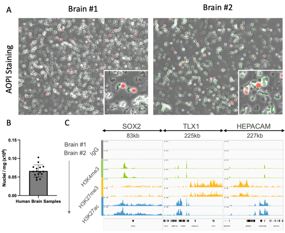
Figure 1. A) Fresh frozen human brain nuclei dissociated with EpiCypher’s automated tissue protocol and stained with acridine orange / propidium iodide (AOPI). Human brain yielded on average 62,000 nuclei per mg of tissue. B) Individual and average yield (in nuclei/mg starting tissue) for 16 human brain tissue samples processed by EpiCypher. C) Example IGV tracks of CUT&RUN assay performed with 500,000 brain nuclei as input. Tracks show enrichment at both neuron and glial associated genes.
Selected brain publications*:
Butts, J et al. A single-cell transcriptomic map of the developing Atoh1 lineage identifies neural fate decisions and neuronal diversity in the hindbrain. Developmental Cell 59, 2171–2188.e7 (2024).
Yamazaki, Shintaro et al. Heat Shock Factor 1 Governs Sleep-Wake Cycles Across Species. bioRxiv 2024.11.15.623879 (2024).
Parmar, Aditya et al. Developmentally dynamic chromatin state at loci regulating organ crosstalk by remote sensing and signaling. Epigenetics Chromatin 18, 82 (2025).
Colon
Nuclei dissociation from frozen colon tissue typically creates minimal debris and is compatible with CUTANA assays without any additional clean-up steps.
Important tips:
Once nuclei are bound to ConA beads, avoid pipetting up and down to resuspend. Instead, distribute clumps by flicking or vortexing your tubes.
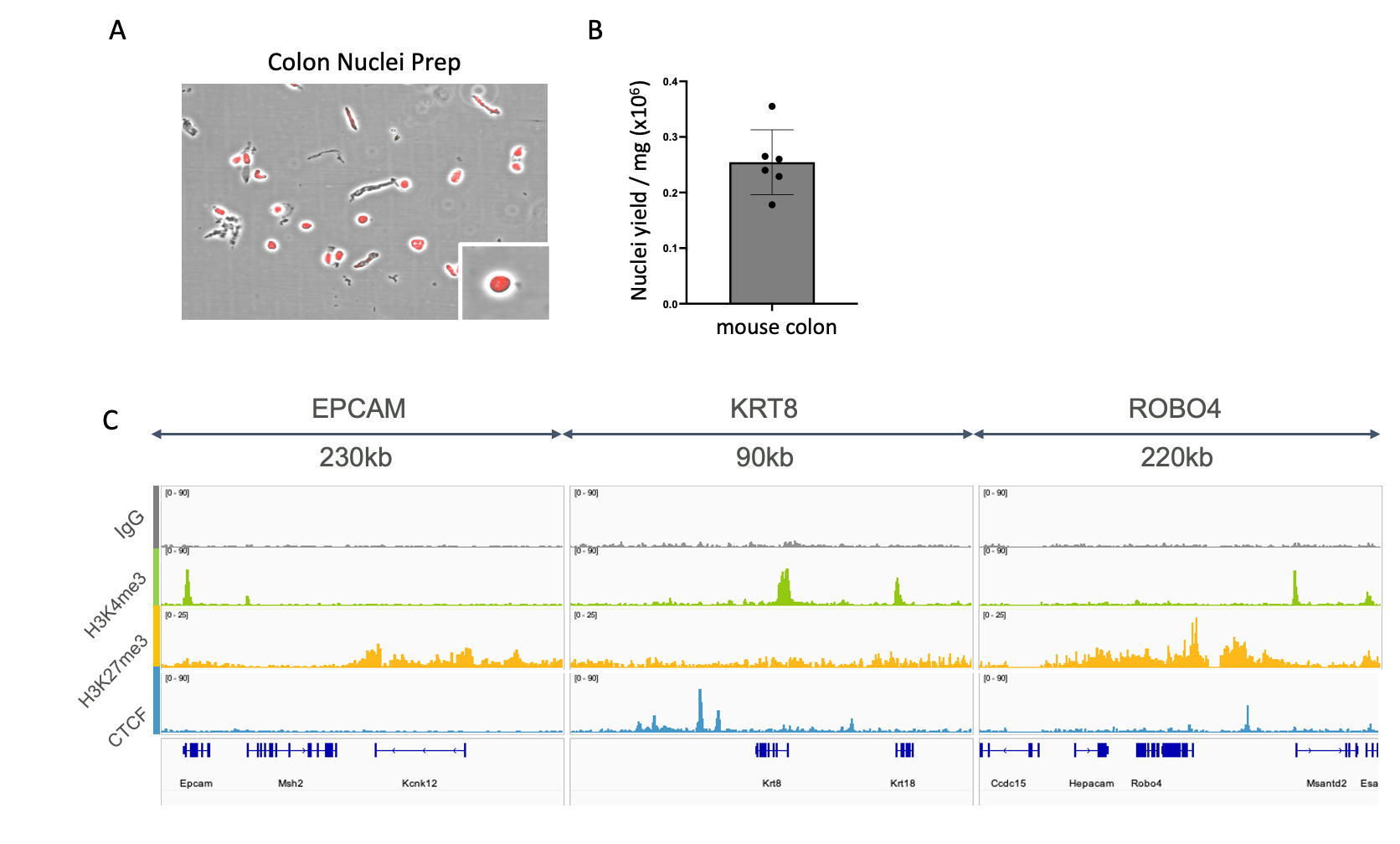
Figure 2. A) Fresh frozen mouse colon nuclei dissociated with EpiCypher’s automated tissue protocol and stained with acridine orange / propidium iodide (AOPI). B) Individual and average yield (in nuclei/mg starting tissue) for six mouse colon tissue samples processed by EpiCypher. Mouse colon yielded on average 250,000 nuclei per mg of tissue. C) Example IGV tracks of CUT&RUN assay performed with 500,000 colon nuclei as input. Tracks show enrichment at colon associated genes.
Selected colon publications*:
Rucli, Sofia et al. Functional genomic profiling of O-GlcNAc reveals its context-specific interplay with RNA polymerase II. Genome Biol 26, 69 (2025).
Janssens, Derek H et al. Automated CUT&Tag profiling of chromatin heterogeneity in mixed-lineage leukemia. Nat Genet 53, 1586–1596 (2021).
Liver
Frozen liver tissue can be challenging to work with, and we have experienced high levels of contaminating high molecular weight gDNA during CUT&RUN. Thus, it is important to minimize the amount of this non-specific DNA going into library prep.
Important tips:
Perform two additional PBS wash steps at the end of your nuclei isolation workflow to remove additional gDNA or lysed nuclei.
Including an IgG negative control is particularly critical when working with liver samples. High DNA recovery in the IgG control following CUT&RUN can indicate that exogenous DNA or contaminating material was carried through from the nuclei preparation. This often serves as an early warning sign of elevated background and reduced signal-to-noise ratio in experimental samples.
Consider adding 5 mM EDTA to your Nuclei Extraction Buffer and PBS washes during nuclei isolation to reduce the impact of endogenous nuclease activity on your sample prep.
Image your nuclei after ConA bead binding and antibody incubation to monitor for nuclei lysis during your CUT&RUN work-flow.
Selected liver publications*:
Parmar, Aditya et al. Polycomb repressive complexes 1 and 2 independently and dynamically regulate euchromatin during cerebellar neurodevelopment. PLoS Genet 21, e1011843 (2025).
Bozukova, Mihaela et al. Aging is associated with increased chromatin accessibility and reduced polymerase pausing in liver. Mol Syst Biol 18, MSB202211002 (2022).
Parmar, Aditya et al. Developmentally dynamic chromatin state at loci regulating organ crosstalk by remote sensing and signaling. Epigenetics Chromatin 18, 82 (2025).
*Important note: EpiCypher has not independently tested the methods used in these publications; they are provided here to complement the guidelines we offer for these sample types. We would recommend beginning with EpiCypher’s tips for your sample type and incorporating aspects of the sample prep given in these publications as necessary.
Don’t see your tissue type here? Check out our searchable publication database and our general tissue dissociation article.