Both fresh and frozen tissue samples can be used as input for CUTANATM workflows. Fresh tissue samples must be dissociated into a single cell suspension using an optimized enzymatic protocol that minimizes cell death and damage. Frozen tissue samples can be prepared by snap freezing biopsies >1cm3 pieces in liquid nitrogen and storing at -80℃ prior to performing a nuclei isolation protocol. Looking for detailed protocols on fresh or frozen tissue dissociation? See this article for automated dissociation (recommended) and this article for manual dissociation.
Effective tissue dissociation preserves the biological characteristics of the original sample while producing a homogeneous preparation with minimal aggregation, low debris, and intact cells or nuclei. Optimizing tissue dissociation is essential because the quality of the starting preparation directly influences downstream assay performance. Poor tissue processing can reduce cell or nuclei recovery, damage fragile cell populations, increase technical variability, and introduce sample preparation artifacts that complicate downstream data interpretation.
Factors that influence the success of tissue dissociation:
Tissue composition: Soft tissues often dissociate readily, whereas fibrotic, connective, adipose, cartilage, or extracellular matrix-rich tissues may require additional optimization.
Sample preservation: While fresh tissues can be used to generate viable single-cell suspensions that can be readily paired with cell isolation techniques, for snap frozen tissues it is better to proceed directly to nuclei isolation.
Desired cell population: Different dissociation methods may preferentially recover or lose certain cell populations, so protocol selection should be guided by experimental goals whenever possible.
Key considerations when dissociating fresh or frozen tissue:
Minimize cell or nuclei aggregation.
Reduce carryover of debris and extracellular material.
Use gentle dissociation methods whenever possible.
Avoid excessive mechanical disruption that may damage chromatin integrity.
For nuclei dissociation from frozen tissue, keep samples cold during processing to preserve sample quality.
When using enzymatic digestion for cell dissociation from fresh tissue, minimize the amount of time the tissue spends exposed to the enzyme. Tissues are highly heterogenous and fragile subpopulations may be preferentially overdigested by enzymatic methods, which introduces bias into your experiment.
Nuclei Isolation from Frozen Tissue
Frozen tissue dissociation can be performed using either manual or automated workflows. We recommend using either a biopulverizor that has been cooled with liquid nitrogen for manual processing, or the GentleMACS® Tissue Dissociator from Miltenyi for automated processing. Using an automated workflow enables higher-throughput processing and consistency, while also resulting in better nuclei yields.
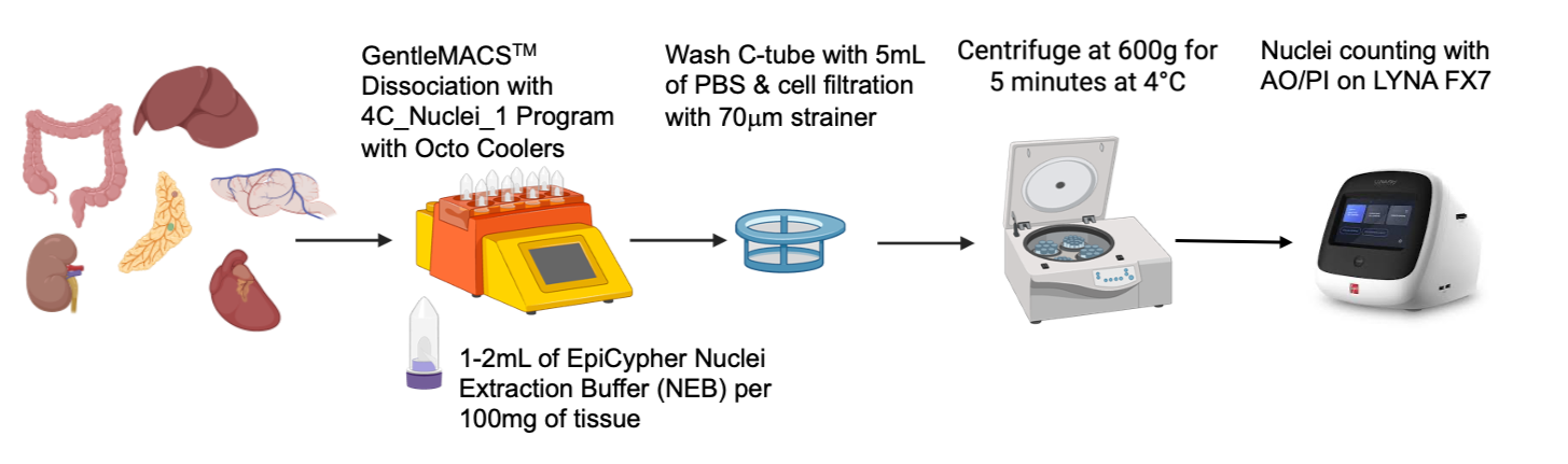
Figure 1. Frozen tissue dissociation schematic using the Miltenyi GentleMACSTM.
Fresh-Frozen Tissue Considerations
Tissue quality, freezing method, storage conditions, and tissue type can all influence nuclei recovery and may require protocol optimization. When processing frozen tissues:
Maintain samples at cold temperatures at all times.
Avoid freeze-thaw cycles.
Use gentle pipetting to preserve nuclei integrity.
Frozen tissue quality can vary substantially between sample types, so pilot experiments may be beneficial when working with a new tissue type.
Evaluate Sample Quality Before Proceeding
Prior to assay setup, it is important to assess sample quality by microscopy and perform nuclei counting. Due to the variability of shape and the presence of debris that often occurs in nuclei derived from tissue, we recommend using a fluorescence counting strategy for this type of sample input. It is still possible to use trypan blue, but quantification can be complicated by the presence of stained debris.
A high-quality preparation of nuclei should exhibit:
Intact morphology
Even dispersion
Minimal aggregation
Low debris levels
If excessive aggregation, debris, or damaged cells or nuclei are observed upon staining, additional optimization of the dissociation protocol may be necessary before proceeding. Sample quality at this stage can significantly impact downstream assay performance and data quality.

Figure 2. Various dissociated mouse tissue samples stained with AOPI. AOPI-stained nuclei dissociated from various mouse tissues. Propidium iodide (red) readily stains nuclei, while Acridine Orange (green) stains various types of debris (especially the myelin prevalent in brain tissue).