Using fluorescent markers for nuclei counting is beneficial when working with tissue, to improve accuracy and assist in distinguishing nuclei from debris. Fluorescent markers also enable automated counting software to give reliable estimates of nuclei quantity and quality. Common markers include DAPI, Hoechst, PI, SYTOX Green, DRAQ5, and DRAQ. Since each of these markers is excited at a different wavelength, your choice of marker should depend on compatibility with your microscope or counting system. Below is a description of EpiCypher’s preferred fluorescent marker, propidium iodide, and a protocol for its use in sample prep for CUTANATM assays.
Propidium Iodide (PI)
PI is a red-fluorescent dye that intercalates into nucleic acids (excitation/emission: 535/617 nm) and is widely used as a counterstain in flow cytometry and microscopy. Because it's excluded by intact, viable cell membranes but readily penetrates compromised ones, PI effectively marks dead cells and nuclei. This makes it especially useful for quantifying extracted nuclei from tissue samples, which tend to have more heterogeneous morphology than nuclei from cultured cells.
PI is often paired with Acridine Orange (AO), which stains live cells as well as various debris commonly present in tissue dissociation preps, thus allowing you to distinguish nuclei from both live cells and debris in a single view.
Important note: Propidium iodide is a potential mutagen, so appropriate safety measures should be taken when working with it.
Nuclei Staining Protocol
Note: Always allow the propidium iodide to equilibrate to room temperature before using.
Prepare a 1 mg/mL (20X) stock solution of prodidium iodide in sterile deionized water for long-term storage (stable for 6 months protected from light at 4 degrees).
Create a working solution by diluting 5 uL of 20X propidium iodide with 95uL of sterile PBS.
Add 10uL of propidium iodide to 10 uL of nuclei suspension.
View using a fluorescence microscope or fluorescence-compatible cell counter, such as the LUNA FX7 or Countess FL3.
Dead cells and nuclei will stain propidium iodide positive (appearing red fluorescent).
Extracellular matrix and debris will remain unstained.
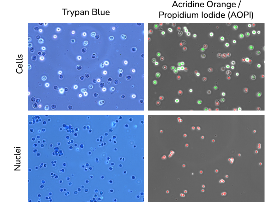
Figure 1. Comparison of cells and nuclei stained with both trypan blue and AOPI. Top panels: cells stained with both trypan blue and AOPI staining. Trypan blue and propidium iodide both stain dead or dying cells with compromised membranes. With trypan blue. live cells appear brighter than dead cells, while living and dead cells are easily distinguished from one another in the propidium iodide sample when acridine orange is added (which stains dead/dying cells green). Bottom panels: extracted nuclei stained with either trypan blue or propidium iodide (red). All nuclei should be permeable to these stains.

Figure 2. Various dissociated mouse tissue samples stained with AOPI.