This article is specific to Fiber-seq. For nuclei extraction for use in CUT&RUN-based assays or CUT&Tag, see this article.
Important Notes
Nuclei Extraction Buffer is prepared fresh on the day of nuclei harvest.
Omission of protease inhibitor in the final Nuclei Extraction Buffer is purposeful, as protease inhibitors are not used in Fiber-seq.
The Pre-Nuclei Extraction Buffer and prepared Nuclei Extraction Buffer are viscous; pay careful attention to preparation notes for both to ensure that they are adequately mixed.
Good sample prep is critical for Fiber-seq. This protocol assumes that you have already harvested the cells you will use to extract nuclei, but is dependent on high-quality starting material.
Note that this protocol is designed for fresh, native human suspension cells.
Materials Needed
Reagent | Source |
|---|---|
Pre-Nuclei Extraction Buffer | |
1M Spermidine | |
Phosphate Buffered Saline (PBS) | Any vendor |
0.4% Trypan Blue | Invitrogen T10282 |
Brightfield or phase microscope + hemacytometer slides | Any vendor |
Nuclei Prep Protocol
1. Prepare 200 µL/reaction Nuclei Extraction Buffer according to the below recipe (Note: After spermidine is added, store at 4ºC for up to 1 week).
Nuclei Extraction (NE) Buffer:
Pre-NE Buffer
0.5 mM Spermidine
2. Count cells, confirm expected morphology, and determine cell viability (as in this article). Ideally, cells should be at least 80% viable, unclumped and with minimal cell lysis or debris. However, ideal viability may vary by sample type, treatments, or processing conditions. The goal is to harvest cells with good integrity and minimal lysis.
3. Transfer 2,000,000 cells per reaction (plus 10% excess if possible) to a 15 ml tube.
4. Spin cells 600 x g for 3 min at room temperature (RT). Aspirate supernatant and resuspend cells in 500 µL PBS to wash once.
5. Transfer cells in 500µL PBS to a 1.5 mL tube.
6. Spin cells 600 x g for 3 min at room temperature (RT). Remove supernatant and resuspend cells in 200 µL per reaction cold Nuclei Extraction Buffer.
7. Incubate for 10 min on ice.
8. Spin 600 x g for 3 min at 4ºC. Pipette to remove supernatant. The pellet should change from pale yellow (cells) to a white, fluffy pellet (nuclei).
9. Gently resuspend nuclei in 75 µL per reaction cold 1X Reaction Buffer.
10. Take a 10 µL aliquot to examine nuclear integrity by trypan blue staining. Isolated nuclei will stain trypan blue positive and should have clear borders and minimal debris. Intact cells will be bright white and round (see Figure 1). For accurate nuclei counts, record "dead" cell numbers on an automated cell counter or manually count blue stained nuclei. Nuclei should be >95% trypan blue positive, unclumped, and show minimal lysis.
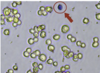
Figure 1. Healthy cells do not stain with trypan blue ( 80% alive). The red arrow indicates a dead cell.