Fluorescence-activated cell sorting (FACS) and magnetic-activated cell sorting (MACS) are compatible with CUT&RUN and CUT&Tag workflows. In addition to isolating cells with specific markers, these methods can remove unwanted debris and dead cells from your sample, potentially improving CUTANA™ assay signal. However, sorting can reduce sample yield and may introduce stress or damage, particularly in fragile cell types. Because CUT&RUN and CUT&Tag require intact membranes for ConA bead binding, gentle sample handling is important. See Figure 1 below for an example workflow incorporating cell sorting into CUTANA sample preparation.
Tips for successful FACS & MACS experiments:
Use gentle sorting conditions. These include: selecting a gentle cell sorter, using ≥ 100µm nozzle size to minimize PSI, and reducing the sort flow rate to minimize shear stress.
Keep all samples on ice before and after sorting.
Include a viability dye, such as propidium iodide (PI) or 7-AAD, to exclude dead or dying cells from your sample.
Perform a 70µm filtration step just prior to sorting.
Native cell inputs are generally less clumpy and produce higher signal than crosslinked cells or isolated nuclei. If you are having difficulty working with native cells, consider either A) performing a light fixation* (0.1% formaldehyde for 1 minute) prior to sorting or B) performing a nuclei extraction post-sort.
For an example of CUT&RUN following FACS, check out the ImmGen data repository, generated in collaboration with EpiCypher. This dataset demonstrates successful CUT&RUN profiling using lightly fixed, cryopreserved nuclei following FACS sorting.
Important Note: Intracellular staining of transcription factors or chromatin-associated proteins is not recommended for CUTANA™ workflows. Intracellular stains may be recognized by pAG-MNase or pAG-Tn5, potentially interfering with assay performance.
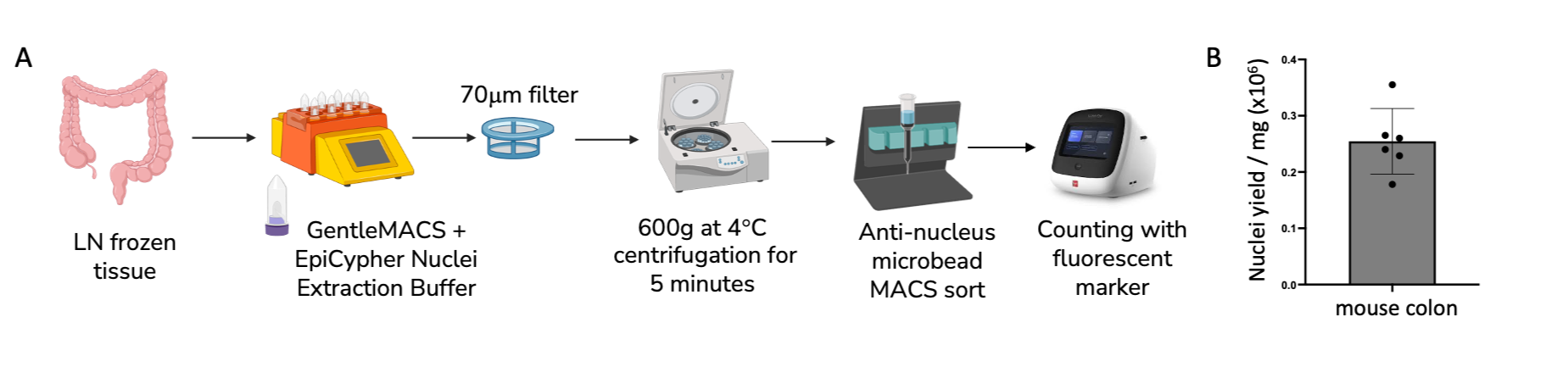
Figure 1. A) Frozen tissue dissociation workflow with anti-nucleus MACS sorting and fluorescent nuclei counting. B) Quantification of average nuclei yield per mg of frozen mouse colon tissue.
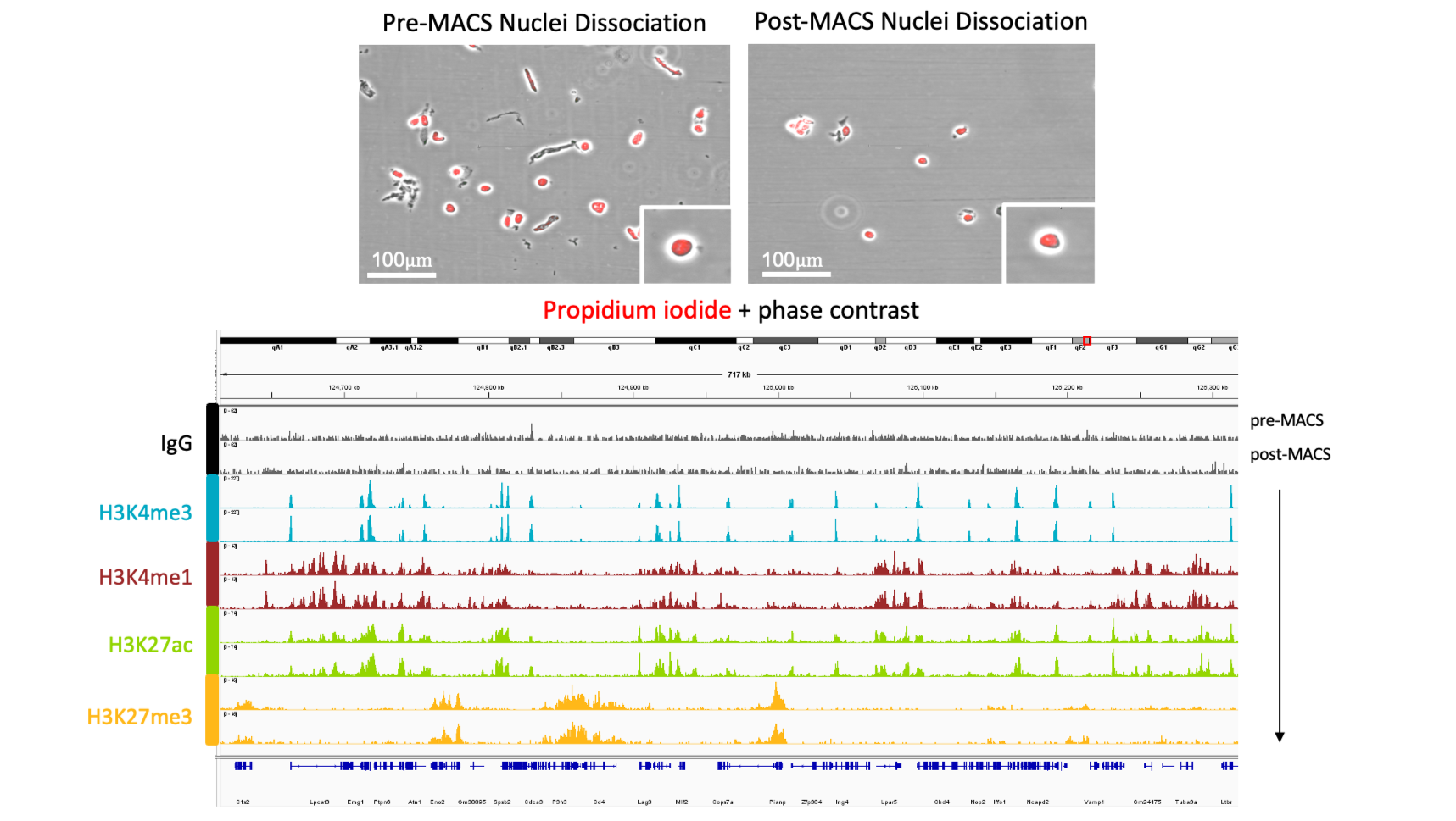
Figure 2. A) Brightfield images of mouse colon nuclei stained with propidium iodide before and after MACS sorting. B) IGV tracks demonstrating the compaitibility of MACS micro-beads with CUTANA genomics workflows.
*For EpiCypher’s CUT&RUN crosslinking protocol, see this article. For our CUT&Tag crosslinking protocol, see this article.