This article also applies to meCUT&RUN and Multiomic CUT&RUN.
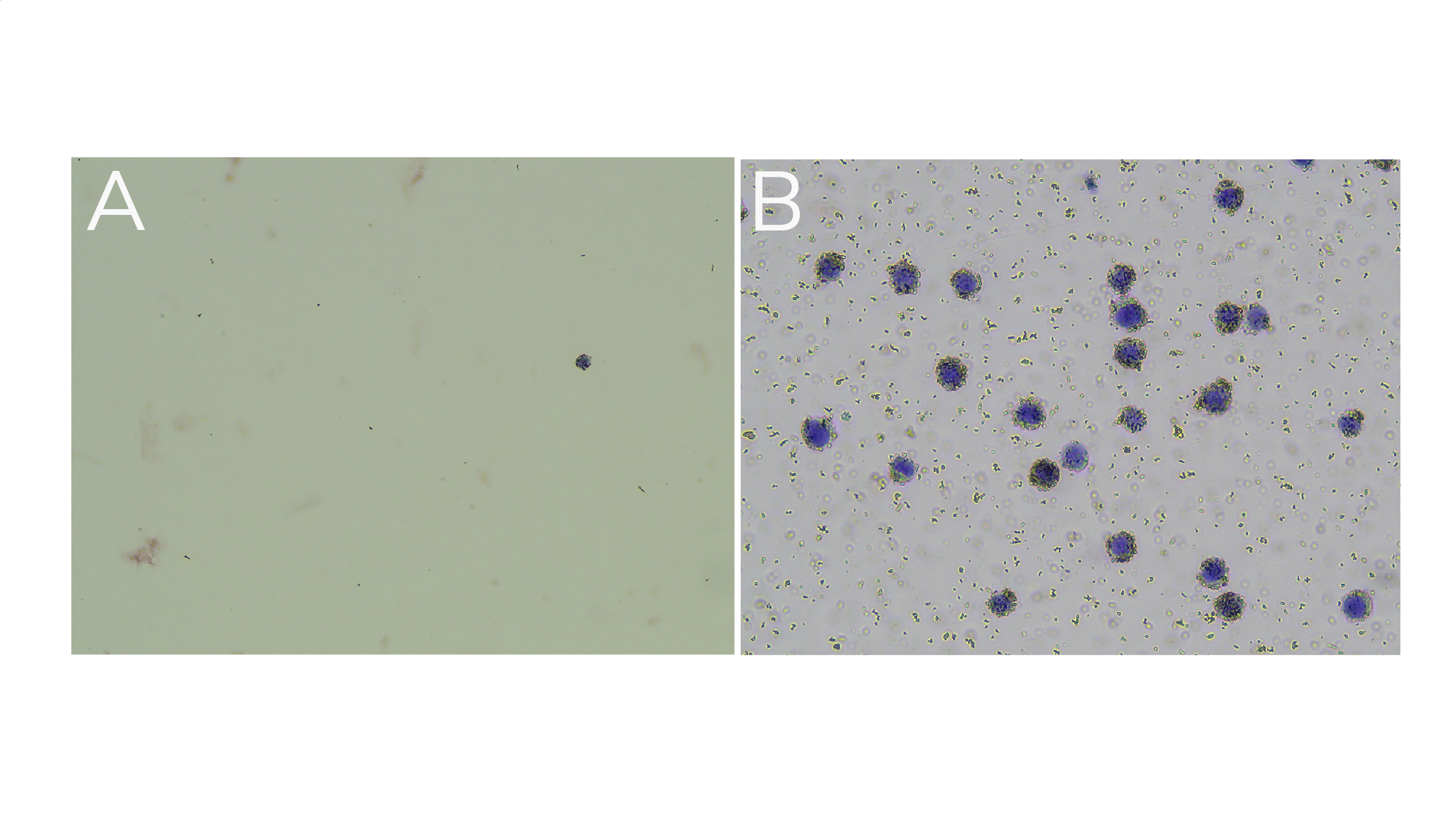
To know if ConA bead binding was successful, we recommend performing Trypan Blue staining following bead binding steps. Save an aliquot of supernatant after ConA bead binding and an aliquot of the cell-bead reaction slurry, and perform Trypan Blue staining for each sample as described. Successful ConA bead binding is indicated when:
The supernatant, or unbound fraction, contains few cells/nuclei (Figure 1A).
The cell-bead reaction slurry contains permeabilized, Trypan Blue positive cells surrounded by ConA beads (Figure 1B).
Should you not observe this, ensure:
Count/integrity of cells before binding to ConA beads.
ConA beads were never frozen.
Cells/nuclei were not clumped.
Beads did not become clumped or dried out.
All buffers were correctly prepared.

Figure 1. (A) Supernatant from ConA bead prep shows few cells/nuclei, whereas (B) cell-bead slurry contains permeabilized (Trypan Blue positive) cells/nuclei surrounded by beads (brown spheres).